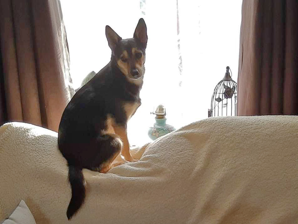
Say hello to Bee, a lovely small crossbreed who loves chasing around and playing in the field.
Unfortunately, Bee got in a bit of a pickle when she ruptured the Cranial Cruciate Ligament in her left stifle (knee joint). She promptly underwent X-rays and surgery, which involved putting a “lateral suture” that would act in a similar manner to the cruciate ligament.
Bee made a full recovery, but it was only 6 months after surgery that trouble started again for her. This time, she damaged the cruciate in the right stifle. It is common for dogs to have problems on both sides within about 18 months from one another.
Things for Bee seemed to be worse this time around because she was also diagnosed with Luxating Patella (slipping of the knee cap), which predisposes to and complicates Cruciate ligament disease.
So, it was clear that Bee had two problems with her right knee (cruciate ligament rupture and slipping of the knee cap), and both needed to be addressed surgically. This is an uncommon situation and requires a good understanding of both conditions, the mechanics of the knee, and good surgical skills.
Bee was internally referred to Luciano, our certificate holder in Small Animal Surgery, for assessment and surgical planning. Soon after, she was scheduled for X-rays and surgery as required. Often, in these cases, the final decision about the best surgical interventions can only be made after the X-rays.
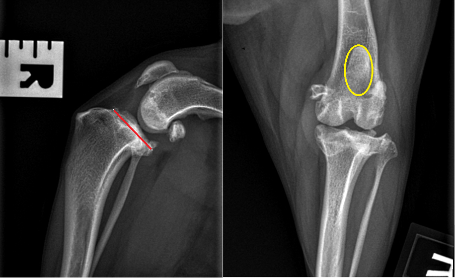
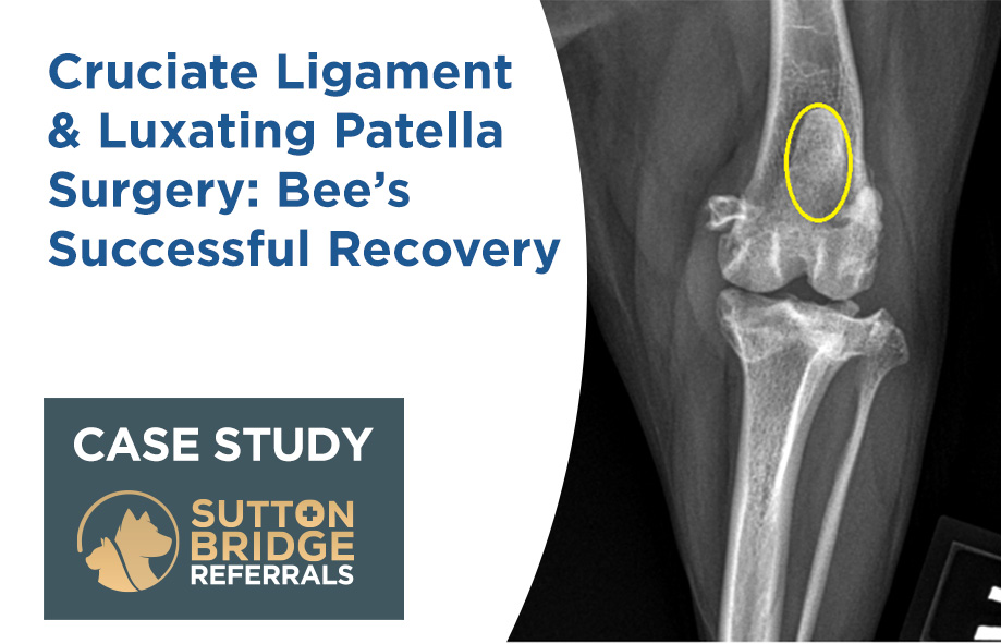
These are Bee’s X-rays. The “red line” shows a steep angle of the tibia (shin bone), which is often found in dogs with cruciate rupture, and the yellow circle around the knee cap.
In agreement with Bee’s Owner, two surgical procedures were carried out in order to correct both problems.
Firstly, the knee joint was inspected, and it was debrided and lavaged. Then, a Wedge Recession Trochleoplasty was performed with a little modification to allow the knee cap to slide and stay in the groove of the femur (thigh bone).
Secondly, a Closing Wedge TPLO for cruciate rupture was carried out. This procedure involves cutting a wedge out of the tibia, re-aligning the bone and fixing it with a plate and screws. This technique also tightens the knee cap tendon, helping address the Luxating Patella problem.
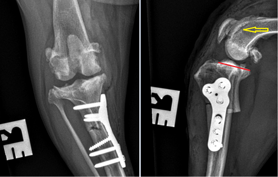
On these post-op X-rays, you can see the implants with the bone cut, the red line showing the bone re-alignment, and the yellow arrow showing where the groove was deepened with the wedge recession trochleoplasty.
Bee was sent home on a course of medications and a rehabilitation plan to follow.
Six weeks later, she made a full recovery and was back in the fields chasing and playing!
We wish Bee a happy life and we are grateful for the attention and care that her owner gave her.
